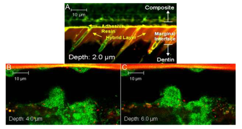
Figure 5:

Confocal images of as prepared (A) and 90-day SHSE degraded (B and C) restoration margins, at various z-depths from the restoration margin edge, infiltrated with live S. mutans (stained green) [16]. Resin adhesive appears at the top of the gap, with dentin on the bottom.
