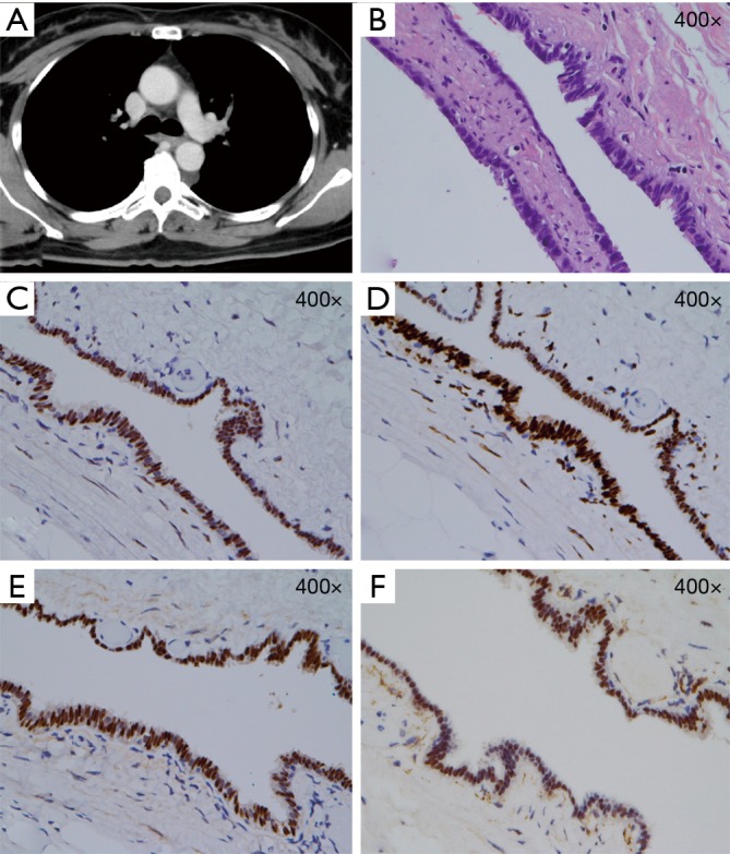
Figure 1.

CT and histological results. (A) Chest CT scan revealed a homogenous soft tissue lesion in the left paravertebral area. (B) Hematoxylin-eosin stain. (C,D,E,F) Immunohistochemical stains for estrogen and progesterone receptors, paired box gene 8, and Wilms’ tumor protein 1.
