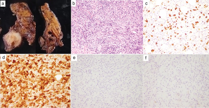
Figure 4.

(a) A macroscopic view of the completely resected mass with a smooth surface, combined with multilocular cysts. (b) Lymphoplasmacytic infiltration and storiform fibrosis with lymphoid follicles on hematoxylin and eosin staining. Immunohistochemical staining for (c) immunoglobulin G4 (IgG4) and (d) IgG. The IgG4/IgG‐positive cell ratio was 60%. (e) Placental alkaline phosphatase and (f) c‐kit staining of the tumor were negative.
