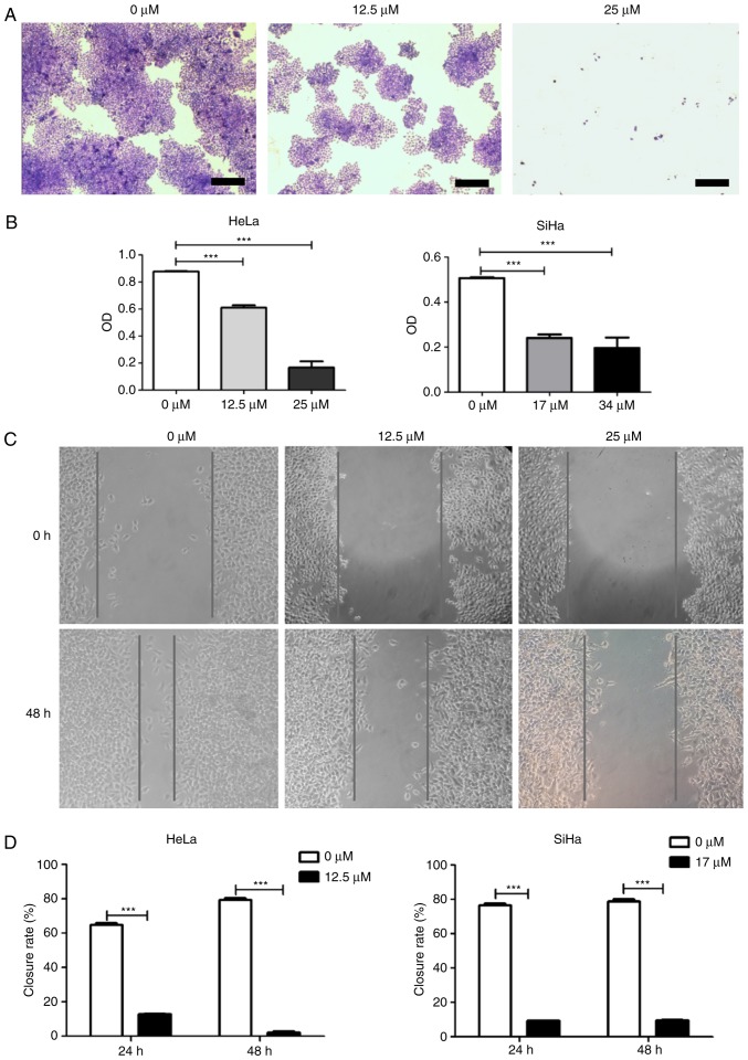
Figure 2.
Colony formation and wound healing assay. (A) Colony formation assay. HeLa or SiHa cells were treated with icaritin for 8 days and stained with crystal violet (representative images of HeLa cells; scale bar, 200 µm). (B) Quantification of colony formation assay. Crystal violet-stained cells were dissolved in 70% alcohol, and absorbance at 595 nm (O.D.) was measured using a microplate reader and presented as mean ± standard error from three independent experiments (***P<0.001). (C) Wound healing assay. Representative images of HeLa cells. (D) Quantification of wound healing assay. Closure rate was defined as follows: (original wound size-new wound size)/original wound size ×100 (n=3, mean ± standard error; ***P<0.001). O.D., optical density.

