
Figure 4.
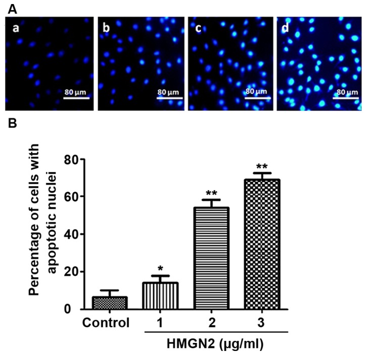
Detection of cell apoptosis via Hoechst staining; (A) image under microscope, bar, 80 µm; concentrations of HMGN2 in (A) a, control; b, 1 µg/ml; c, 2 µg/ml; d, 3 µg/ml. (B) Examination of apoptosis by Hoechst assay. Compared with the control group, HMGN2 in different doses induced cell apoptosis. *P<0.05, compared with control group; **P<0.01, compared with control group.