Figure 1.
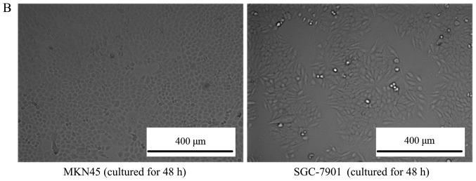
Morphology of cultured cells. (A) The morphology of mesenchymal stem cells observed by inverted phase-contrast microscope (magnification, ×100). The left picture is the MSCs cultured for 12 h, where all the cells adhered to the bottom of the culture dish and cells were observed to be round. For the middle picture, which depicts MSCs cultured for 24 h, the cells began to grow in a fibroblast-like manner, and the right picture is the MSCs cultured for 48 h, depicting that ~80% of the cells had fused together. (B) The morphology of MKN45 and SGC-7901 cells observed by inverted phase-contrast microscope (magnification, ×100). The left picture is the MKN-45 cells cultured for 48 h, where all the cells grew in a triangle or quadrangle manner. The right picture is the SGC-7901 cells cultured for 48 h, where the cells grew in classical short shuttle-like manner. MSCs, mesenchymal stem cells.


