Figure 1.
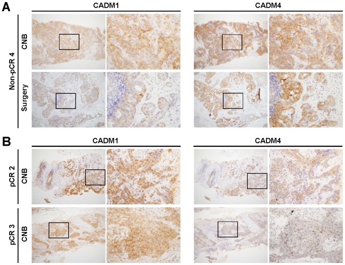
Representative images of CADM1 and CADM4 immunohistochemical staining of CNB and surgical specimens. (A) A non-pCR case with both positive CADM1 and CADM4 staining in CNB and surgical specimens. (B) A pCR case (pCR 2) with positive CADM1 and positive CADM4 staining in CNB (upper four panels). A pCR case (pCR 3) with positive CADM1 and negative CADM4 staining in CNB (lower four panels). Left low magnification (×100) and right high magnification (×400) panels are shown. A high magnification image (×400) of the region in the black rectangle of the left panel is presented in the right panel. pCR, pathological complete response, CNB; core needle biopsy; CADM, cell adhesion molecule.

