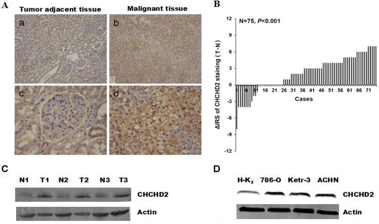
Figure 1.
CHCHD2 protein expression was determined in RCC tissues and cells by immunohistochemical staining and western blot analysis. (A) Representative images revealed CHCHD2 immunohistochemical staining in (b and d) RCC maligant tissues and (a and c) tumor adjacent tissues, which were taken at different magnifications (Top panel, ×100; bottom panel, ×400). (B) The distribution of the difference in CHCHD2 staining (ΔIRS=IRST-IRSN). Immunoreactivity score (IRS) of CHCHD2 staining was available from 75 pairs of tissues. P-values were calculated with the χ2 test. (C) Whole-cell protein extracts were further prepared from three paired tumor adjacent normal renal tissues and RCC tissues. The CHCHD2 protein level was determined by western blot analysis. (D) In contrast to normal renal HK-2 cell, the CHCHD2 expression is increased in RCC cell lines. RCC, renal cell carcinoma; CHCHD2, coiled-coil-helix-coiled-coil-helix domain-containing protein 2; N, normal renal tissues; T, RCC tissues; IRS, immunoreactivity score.

