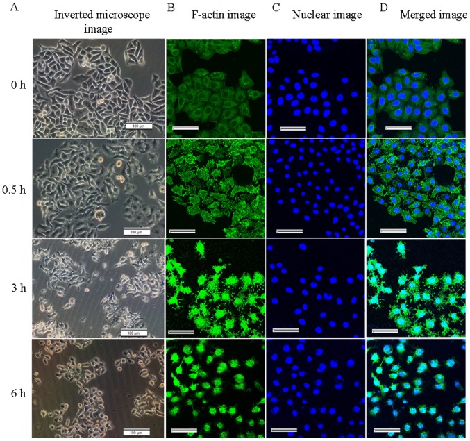
Figure 1.
Effect of cytochalasin B (CB) on cell morphology and the F-actin cytoskeleton at different time-points. (A) Optical micrographs. Scale bars, 100 µm. Untreated cells exhibited normal morphology, while the CB-treated cells gradually shrank, rounded up, even became detached from the substrate and floated. (B) In addition, F-actin was disrupted in the treated cells, and the short or punctate actin fragments (green fluorescence) occupied the background of the images. (C) The nucleus (blue fluorescence) appeared irregular at 6 h. (D) Merged images. Scale bars, 50 µm.

