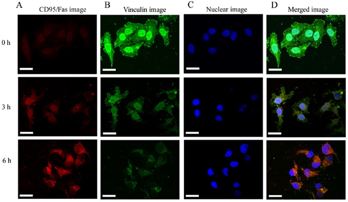
Figure 3.
Immunofluorescence staining of HeLa cells death receptor CD95/Fas and vinculin protein under treatment with cytochalasin B. (A and D) Only a mild activation of CD95/Fas was observed, indicated by weak red fluorescence at 3 h, whereas the red fluorescence was considerably brighter at 6 h. (B and D) By contrast, in untreated cells, the vinculin staining exhibited bright dot green fluorescence, whereas in the treated cells it was continuously diminished, with only a small amount of green fluorescence surrounding the nucleus at 6 h. (C) The nucleus (blue fluorescence) appeared irregular at 6 h. Scale bars, 20 µm.

