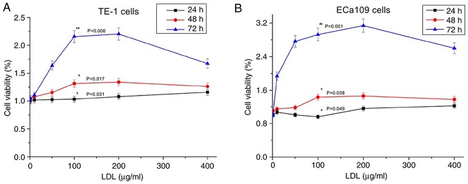
Figure 2.
Effect of LDL on TE-1 and ECa109 esophageal cancer cell viability. Cells were incubated with various concentrations of LDL (0–400 µg/ml) for 24 h (black line), 48 h (red line) and 72 h (blue line). (A) The TE-1 proliferation rate increased with the treatment time and with a concentration <200 µg/ml; the proliferation rate decreased with LDL concentrations between 200 and 400 µg/ml (*P<0.05 vs. 24 h; *P<0.05 vs. 48 h; **P<0.01 vs. 72 h). (B) Similar results were obtained using ECa109 cells (*P<0.05 vs. 24 h; *P<0.05 vs. 48 h; **P<0.01 vs. 72 h). LDL, low-density lipoprotein.

