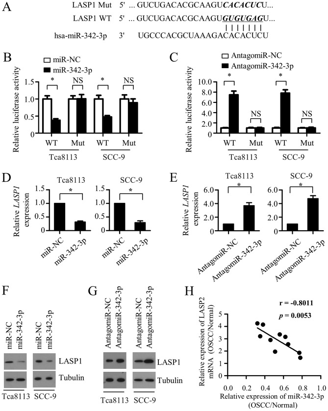
Figure 4.
miR-342-3p directly targets LASP1 in OSCC cells. (A) Predicted miR-342-3p target sequence in the 3′-UTR of LASP1 mRNA, and the WT and mutated versions used in the luciferase reporter plasmids. Luciferase activity data (shown as the mean ± SD of triplicate measurements) of (B) Tca8113 and SCC-9 cells transfected with WT or mutant LASP1 3′-UTR luciferase reporter plasmid along with miR-NC or miR-342-3p, and (C) Tca8113 and SCC-9 cells co-transfected with antagomiR-NC or antagomiR-342-3p and WT or mutant LASP1 3′-UTR luciferase reporter vectors. Quantitative PCR was performed to detect the expression of LASP1 mRNA in Tca8113 and SCC-9 cells transfected with (D) miR-NC or miR-342-3p or (E) antagomiR-NC or antagomiR-342-3p. Expression of LASP1 mRNA was normalized to GAPDH. Results are shown as the mean ± SD of triplicate measurements. (F) Western blotting was used to measure the expression of LASP1 protein in Tca8113 and SCC-9 cells transfected with (F) miR-NC or miR-342-3p or (G) antagomiR-NC or antagomiR-342-3p. α-tubulin was used as a loading control. (H) A statistically significant inverse correlation between miR-342-3p expression and LASP1 mRNA in OSCC vs. normal tissues from the same patient was observed following Pearson's correlation analysis (R=−0.8011; P=0.0053). *P<0.05. miR, microRNA; LASP1, LIM and SH3 protein 1; OSCC, oral squamous cell carcinoma; 3′-UTR, 3′ untranslated region; WT, wild-type; NC, negative control; SD, standard deviation; NS, not significant.

