Figure 2.
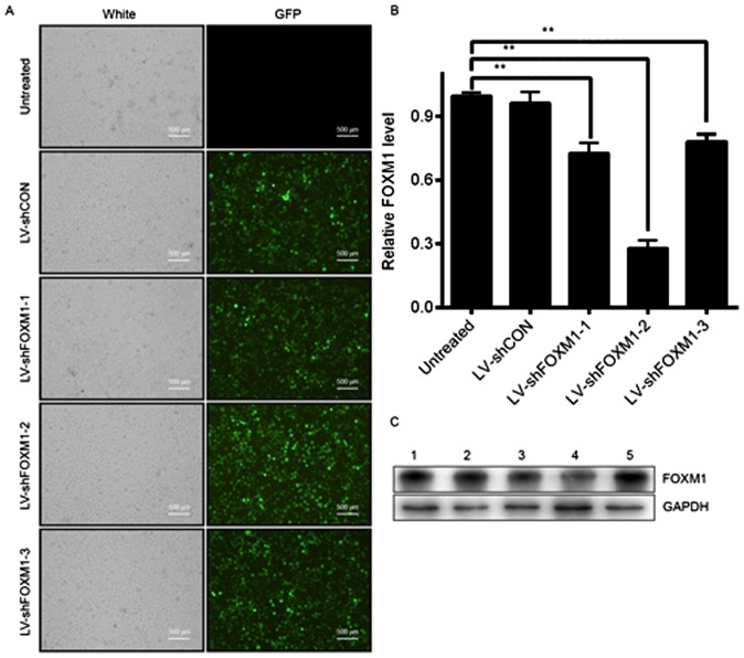
Selection of the optimal LV-shFOXM1 in Tca8113 cells. (A) GFP expression in Tca8113 cells after 48 h of infection, determined under a fluorescence microscope. (B) mRNA level of FOXM1 following lentiviral infection, determined using reverse transcription-quantitative polymerase chain reaction. (C) Protein level of FOXM1 determined using western blot analysis. GADPH was used as the loading control. Lane 1, Tca8113 cells were not infected; lane 2, Tca8113 cells were infected with LV-shCON; lane 3, Tca8113 cells were infected with LV-shFOXM1-1; lane 4, Tca8113 cells were infected with LV-shFOXM1-2; and lane 5, Tca8113 cells were infected with LV-shFOXM1-3. **P<0.01 vs. untreated. LV-shFOXM1, lentivirus-short hairpin RNA Forkhead box M1; CON, control; GFP, green fluorescent protein.

