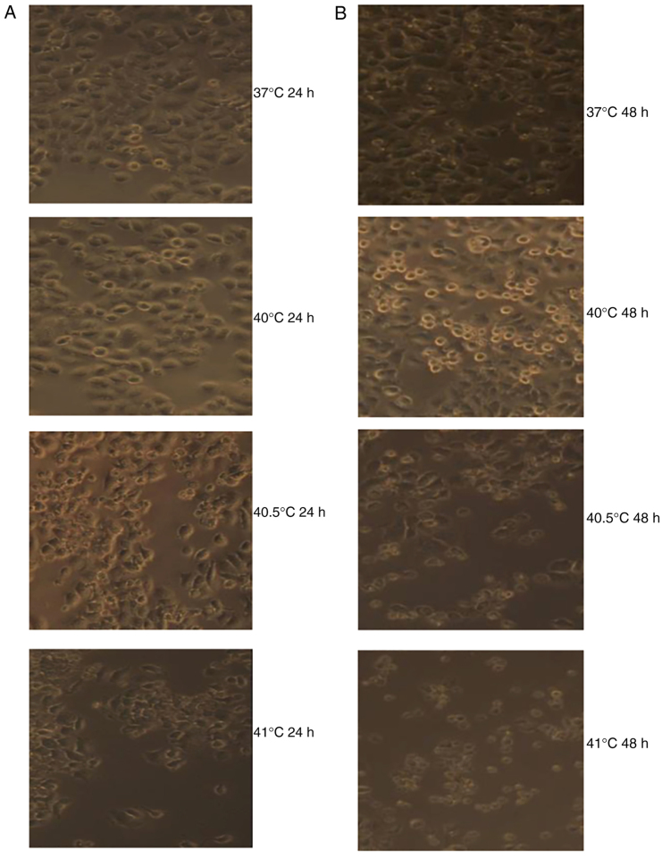
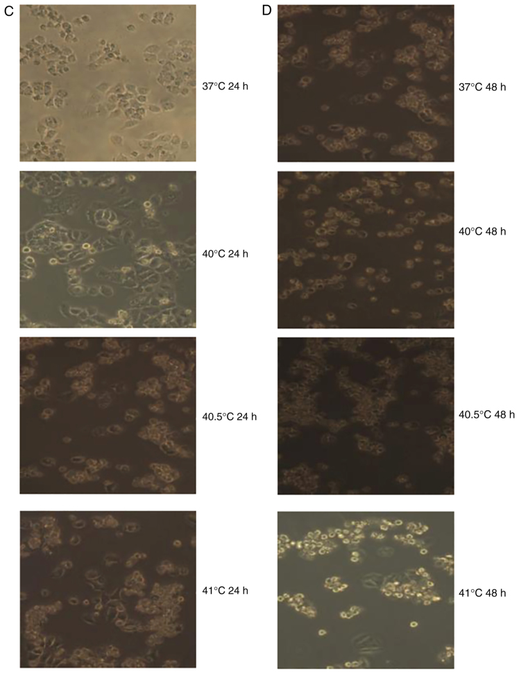
Figure 4.
Photomicrographs displaying the effects of the combination of paclitaxel and mild hyperthermia on MCF-7 cells. (A) MCF-7 cells 24 h post-treatment with 5 µg/ml of paclitaxel and exposure to mild hyperthermia. (B) MCF-7 cells 48 h post-treatment with 5 µg/ml paclitaxel and exposure to mild hyperthermia. (C) MCF-7 cells 24 h post-treatment with 10 µg/ml of paclitaxel and exposure to mild hyperthermia. (D) MCF-7 cells 48 h post-treatment with 10 µg/ml of paclitaxel and exposure to mild hyperthermia. Cultures exposed to hyperthermia at 40°C displayed reduced apoptosis compared with cultures treated with 5 µg/ml paclitaxel alone, while cultures treated with hyperthermia at 40.5 or 41°C displayed greater numbers of dead cells (due to apoptosis or necrosis) compared with cultures treated with 5 or 10 µg/ml or paclitaxel alone. The numbers of dead cells increased with longer time after treatment. The images of 37°C 24 h and 37°C 48 h are the results of paclitaxel (5 or 10 µg/ml) alone. Magnification, ×100.


