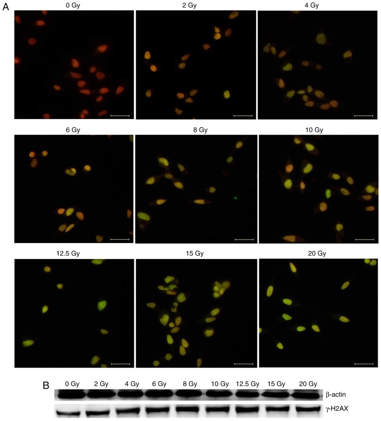
Figure 2.
Radiation-induced DNA damage. (A) DNA double-strand breaks were visualized with immunofluorescent staining for γ-H2AX foci. γ-H2AX stained with green fluorescence (fluorescein isothiocyanate) reflects DSBs and merged with nucleus stained with red fluorescence (propidium iodide). Significant dose-dependent increase in foci number was detected following X-irradiation in HeLa cells. Scale bar, 100 µm. (B) Results of western blot analysis demonstrated the expression of γ-H2AX was increased following irradiation. γ-H2AX, phosphorylated histone H2AX.

