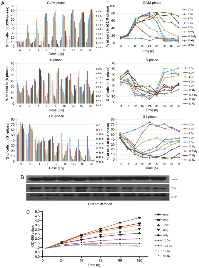
Figure 3.
Cell cycle following irradiation. (A) Cell cycle (G1, S and G2/M) changes were analyzed by flow cytometry at 0, 6, 12, 18, 24, 30, 36, 42 and 48 h after X-ray irradiation using propidium iodide staining. Irradiation of HeLa cells resulted in a notable increase of cells in the G2/M-phase. (B) Results of western blot analysis demonstrated the level of CDK1 and CHK1 in cells following irradiation, the expression of CDK1 markedly decreased following various doses of irradiation and the expression of CHK1 increased following irradiation. (C) Cell proliferation was detected using Cell Counting Kit-8 assays in cells following irradiation. CDK1, cyclin dependent kinase 1; CHK1, checkpoint kinase 1; OD, optical density.

