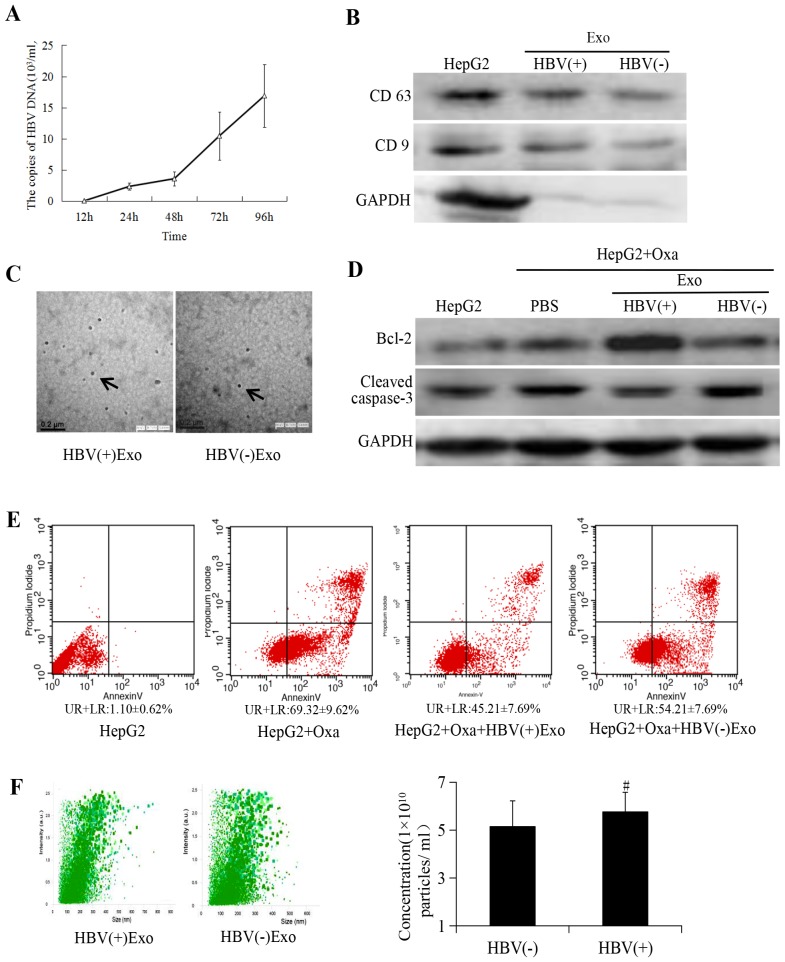
Figure 2.
Exosomes derived from HBV-positive liver cancer cells promote cell chemoresistance. (A) HBV-DNA contents in culture supernatant reached a peaked at 96 h. Exosomes were isolated according to manufacturer's instructions (47). HepG2 cells were treated with HBV-positive serum (HBV-DNA 1×10¹⁰ copies/ml) and HBV-DNA copy of the cell supernatant containing pure DMEM was detected at different times following removal of serum; experiments demonstrated that HBV-DNA copies reached a peaked (16.89±5.02×10³ copies/ml) at 96 h comparing with HBV-DNA copies in 12, 24, 48 h, which provided the basis for subsequent experiments. (B) Exosome markers CD63 and CD9 were assessed by western blotting. GAPDH was used as an internal reference. (C) Exosomes were characterized as round vesicles when examined using transmission electron microscopy (magnification, ×40,000). (D) Detection of cell apoptosis on treatment with 1×10¹⁰ particles of HBV-associated exosomes in the experimental group and an equal amount of particles of non-HBV-associated exosomes in the negative group. (E) Flow cytometry analysis of cell apoptosis. (F) Analysis of size distribution and exosome concentration in purified exosomes using Nanosight technology. No statistically significant differences were indicated between HBV-associated and non-HBV-associated liver cancer cells concerning exosomes concentration. #P>0.05 vs. exosomes from non-HBV-associated liver cancer cells. HBV, hepatitis B virus; Bcl-2, B-cell lymphoma; CD, cluster of differentiation; GAPDH, glyceraldehyde 3-phosphate dehydrogenase; Exo, exosome; Oxa, oxaliplatin; UR, upper right; LR, lower right.

