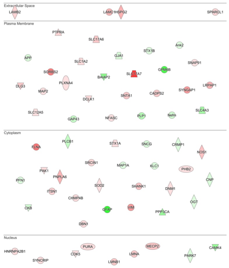
Figure 4.
Drivers of neuronal morphology affected by G-CSF. IPA analysis of the most significantly altered cellular functions following G-CSF treatment revealed that proteins involved in altering neuronal morphology were significantly changed. This diagram shows all significantly regulated proteins predicted to be involved in affecting neuronal morphology, and their corresponding predicted subcellular distribution. Proteins visualized in red are significantly increased, and those in green were significantly decreased.

