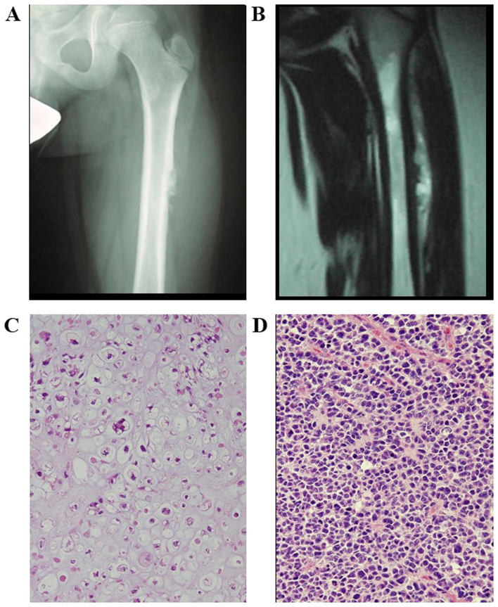
Figure 1.
Case 1. (A) Radiograph of the left proximal femur demonstrating an aggressive lytic bone lesion with spiculated periosteal reaction in the proximal femoral metadiaphysis. (B) Coronal T2-weighted magnetic resonance imaging showing heterogeneous expansive mass, with high signal intensity. (C) Pathological examination confirmed chondroblastic osteosarcoma (hematoxylin and eosin staining; magnification, ×200). (D) Primary cancer: Medulloblastoma of the cerebellar vermis (hematoxylin and eosin staining; magnification, ×200).

