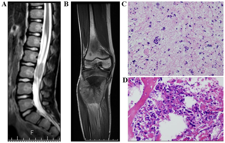
Figure 3.
Case 3. (A) Sagittal T2-weighted magnetic resonance imaging (MRI) showing a heterogeneous, isointense mass. (B) Coronal T2-weighted MRI showing a heterogeneous, hyperintense expansible mass. (C) Primary cancer: Astrocytoma at the intradural region of the L3 level (hematoxylin and eosin staining; magnification, ×200). (D) Pathological examination confirmed osteoblastic osteosarcoma (hematoxylin and eosin staining; magnification, ×200) (d).

