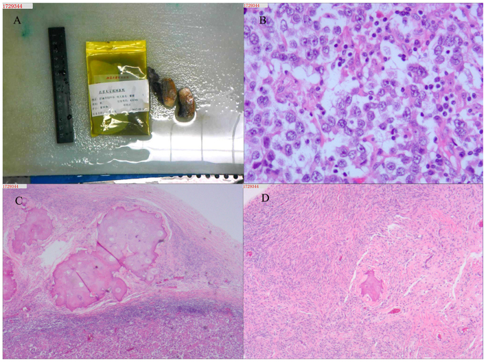
Figure 3.
(A) Gross examination of removed testicular tissue, sized 4.0×2.5×1.2 cm. The cut surface was yellowish-brown and soft, and included a grey mass, slightly harder in texture. The size of the epididymis was 4.0×1.5×1.0 cm. (B) In testicular tissue, spermatogenic cells in the seminiferous tubules were highly atrophic or absent, the basement membrane was significantly thickened, and hyalinization was observed, along with stromal cell proliferation. (C) Ovarian-like tissue was detected in an area of the testicular tissue. (D) In the testicular tissue, more irregularly shaped large cells were arranged in a nodular pattern, with scattered interstitial lymphocytes. Combined with immunohistochemistry, the findings were consistent with seminoma.

