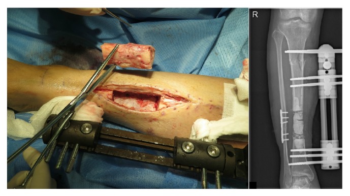
Figure 2.

The left figure was the resection of infected bone segment when radical debridement was conducted. The right figure was the anteroposterior X-ray photograph taken few hours after surgery, and antibiotic-impregnated calcium sulphate was demonstrated to be filled in the bone defect.
