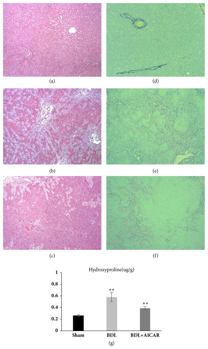
Figure 1.
Hematoxylin & eosin (H&E), sirius red staining and hydroxyproline assay of rats liver tissue. Panels (a) and (d) show normal liver tissue of the H&E and Sirius red staining. Panels (b) and (e) represent BDL liver tissue with large areas of necrosis, inflammatory cell infiltration, and fibrosis. Newly formed bile ducts were also observed in BDL liver. Panels (c) and (f) represent less inflammatory cell infiltration and a decreased number of newly formed bile ducts in the BDL rats with the application of AICAR. Panel (g) shows the hydroxyproline content in three groups. ∗∗ P value <0.05, which was considered to be statistically significant in three groups.

