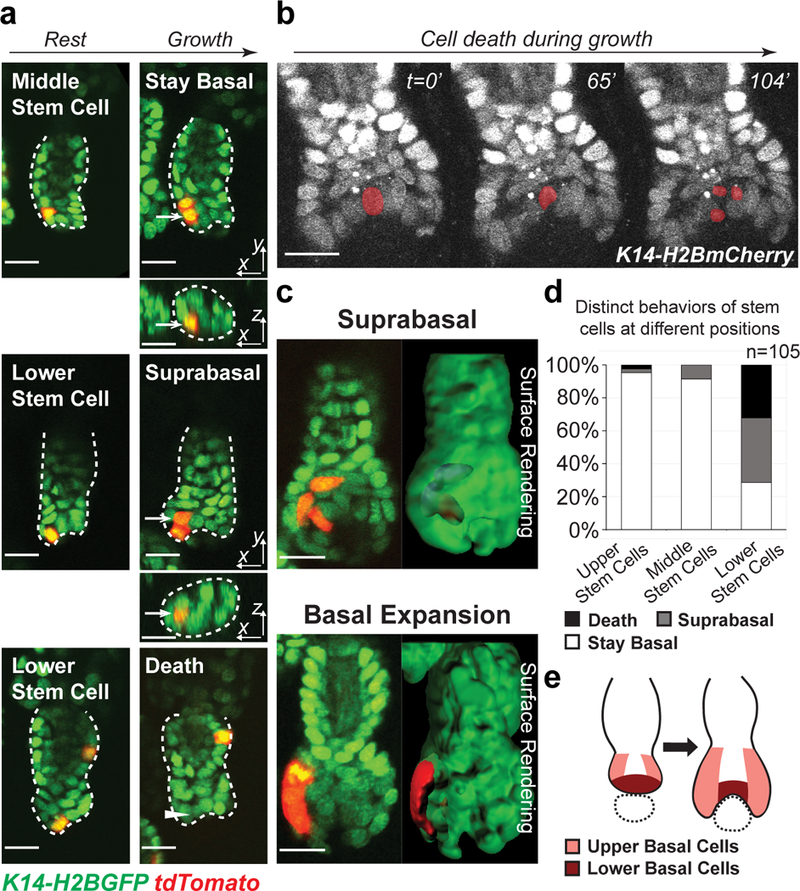
Fig 2. Spatially regulated stem cell behaviors lead to spatially reversed organization of stem cell progeny.

a, Representative examples of lineage tracing of single hair germ stem cells at different positions in early hair follicle growth phase, showing progeny of middle stem cells stayed basal, while progeny of lower stem cells were displaced into the suprabasal layer or underwent cell death. Arrows indicate the stem cell progeny. Arrowhead indicates where the dead stem cell was. Hair follicle epithelium is outlined by dashed lines. The tracking was typically performed from Telogen to Anagen I. Images representative of 105 hair follicles from 6 mice. b, Time-lapse frames showing stem cell death at lower position. The dying cell is pseudo-colored. Images representative of 5 mice. Two independent time-lapses are provided in Supplementary Video 3. c, Single z-planes and 3D surface rendering of hair follicles during lineage tracing showing suprabasal displacement (blue) of cells deriving from lower stem cell (up) and basal expansion of cells deriving from upper stem cell (down). d, Frequency of hair germ stem cells at different positions undergoing cell death, suprabasal displacement and basal expansion. 100% stacked column shows the percentage of hair follicles with a single stem cell labeled at a given position that performed a certain behavior. n=105 hair follicles in 6 mice. e, A schematic showing distinct behaviors of hair germ stem cells at different positions lead to differential basal expansion and mesenchyme encapsulation. Dotted circle indicates mesenchymal dermal papilla. Epithelial nuclei were marked by K14-H2BGFP (green in a and c) or K14-H2BmCherry (white in b). Lgr5-CreER and R26-flox-STOP-tdTomato (red in a and c) were used to induce stem cell labeling. Scale bars, 20 μm.
