
Fig 4. Hair progenitors undergo dynamic relocation and change differentiation outcomes.
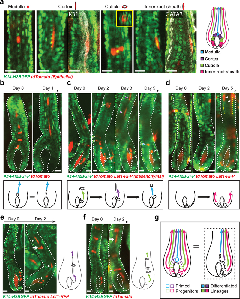
a, Morphologies and positions of differentiated hair lineages. Whole mount staining of cortex marker (K31, white) and inner root sheath (IRS) marker (GATA3, white) confirms the molecular signature of differentiated cells. XZ view of the labeled cuticle cell showing its distinctive ring shape (inset). Images representative of 20 mice. b, Lineage tracing of matrix progenitors and schematic show top progenitors stop self-renewal. Images representative of 44 hair follicles from 11 mice. c, Lineage tracing of matrix progenitors and schematic show lower progenitors are continually relocated upwards and change differentiation outcomes (arrowheads) corresponding to their new positions. Arrowheads with different colors indicate distinct differentiation lineages produced at each time point. Images representative of 149 hair follicles from 17 mice. d, Lineage tracing of outer root sheath (ORS) cells and schematic show ORS cells move into the matrix and produce differentiated lineages (arrowheads). Images representative of 49 hair follicles from 8 mice. Yellow dashed lines indicate the interface between ORS and inner lineages. e, An example of concurrent multi-lineage differentiation of matrix progenitors and schematic showing a single progenitor produced an outer differentiated cell (cuticle cell, arrow) among cortex cells (arrowhead). f, An example of concurrent multi-lineage differentiation of matrix progenitors and schematic showing a single progenitor produced an inner differentiated cell (cortex cell, arrow) among cuticle cells (arrowhead). g, A model of primed progenitors with distinct molecular signatures undergo dynamic relocation and change differentiation outcomes. Epithelial nuclei were marked by K14-H2BGFP (green in a, b, c, d, e and f). Hopx-CreER (a, b, c, e and f) or Lgr5-CreER (b, c, d) in combination with R26-flox-STOP-tdTomato (red) was used to induce cell labeling. The tracking was performed from Telogen (b) or Anagen IIIc (b, c, d, e and f) to Anagen VI. Hair follicle epithelium is outlined by dashed lines (b, c, d, e and f). Mesenchymal dermal papilla was labeled by Lef1-RFP in c, d and e. Asterisk and arrow indicate the original and current progenitor cell position respectively in b, c and d. Scale bars, 20 μm.