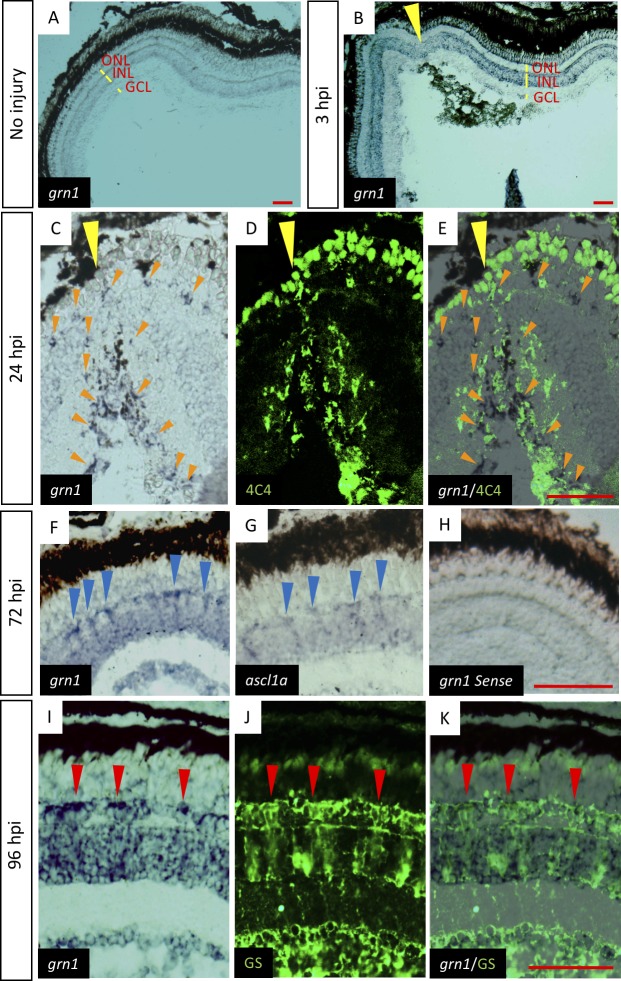
Figure 2.
Alterations in expression of grn1 in retinal cells during retinal regeneration. Time-course in situ hybridization analysis shows that grn1 is expressed in different retinal layers at each time point. In retina, melanin mainly accumulates at the RPE and in situ labeled grn1 mainly localized inner retinal layer. In situ labeling also shows weaker signal than melanin. (A) In situ hybridization of grn1 at uninjured control retina. grn1 is weakly expressed in the INL. (B) In situ hybridization of grn1 at 3 hpi. Yellow arrowheads indicate the injury site. grn1 is expressed in the GCL, photoreceptor cells, and INL. The areas that are very close to the injury site express more grn1, whereas the injury site has lower expression of grn1. (C–E) Retina were stained at 24 hpi. Orange arrowheads indicate expression site of grn1. Yellow arrowheads indicate the injury site. (C) In situ hybridization of grn1. (D) Anti-4C4 antibody staining. (E) Merged image. (F–H) Retina were stained at 72 hpi. Blue arrowheads indicate the strong expression site of grn1 or ascl1a. (F) In situ hybridization of grn1. (G) In situ hybridization of ascl1a. (H) In situ hybridization using grn1 sense probe. (I–K) retina were stained at 96 hpi. Red arrowheads indicate the colocalization of grn1 and glutamine synthase (GS). (I) In situ hybridization of grn1. (J) Anti-GS antibody staining. (K) Merged image. Scale bars: 50 μm.

