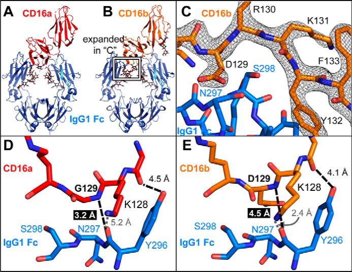
Figure 5.

CD16a binds IgG1 Fc with greater affinity and makes a closer approach to IgG1 Fc through the loop containing residue 129. A and B, the overall complexes formed by CD16a (A) and CD16b (B) with IgG1 Fc are highly similar with a root mean square deviation of 0.499 Å. The protein backbone is shown as ribbons, and N-glycans are shown as sticks. C, the electron density of the CD16b region surrounding Asp-129 is well-resolved at 1.5 σ. D and E, the Gly-129 amide nitrogen atom of CD16a (D) is located 1.3 Å closer to the carbonyl carbon of the IgG1 Fc Asn-297 than the amide nitrogen of Asp-129 from CD16B (E).
