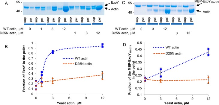
Figure 6.
ExoY and MBP-fusion binding to WT or D25N actin. A, SDS-PAGE analysis of cosedimentation experiments of 2 μg (3 μm) of full-length ExoY and yeast F-actin. B, the fractions of ExoY that cosedimented with F-actin were quantified by densitometry using ImageJ software and were plotted against actin concentrations. Error bars correspond to standard deviations of three independent experiments. C, SDS-PAGE analysis of cosedimentation experiments of 2 μg (3 μm) of fusion protein MBP–ExoY360–378 and yeast F-actin. D, the fractions of the fusion protein MBP–ExoY360–378 that cosedimented with F-actin were quantified by densitometry using ImageJ software and were plotted against actin concentrations. Error bars correspond to standard deviations of two independent experiments. sup, supernatant; pel, pellet.

