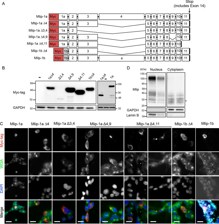
Figure 4.
Cellular localization of MLIP isoforms. A, schematic representation of the different synthetic human Myc-tagged Mlip isoforms used to transfect 293 cells. B, Western blot with Myc tag antibody. GAPDH was used as a loading control. C, immunostaining of 293 cells with Myc tag antibody (red), wheat germ agglutinin (WGA; green), and 4′,6-diamidino-2-phenylindole (DAPI; blue). Scale bars, 20 μm. D, Western blot with MLIP antibody of nuclear and cytoplasmic fractions isolated from MLIP+/+ heart. GAPDH and lamin B were used as cellular fractionation controls. *, cytoplasmic-specific isoform.

