Figure 3.
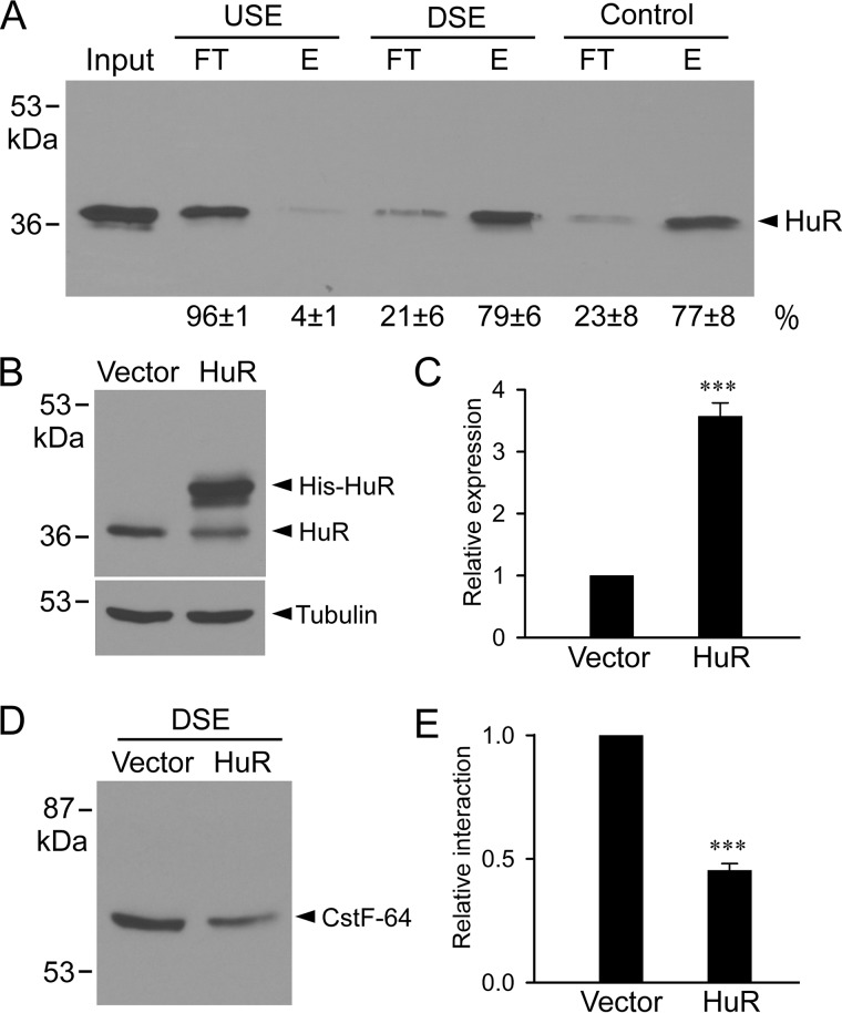
Interaction of HuR with intron 9 poly(A) signal downstream sequence. A, cell lysates from HEK293 cells were incubated with biotinylated and streptavidin bead–bound RNA probes corresponding to the DSE, the USE, or the control HuR-binding sequence from the 3′-UTR of androgen receptor mRNA. The RNA-bound protein fractions (E) and unbound fractions (FT) were analyzed by immunoblotting with anti-HuR antibody. Signals were quantified and expressed as percentage of total (FT + E) protein (n = 4). B, immunoblot analysis of total nuclear extracts from vector or HuR transfected cells. C, histogram showing the significant increase in HuR expression following transfection compared with vector-transfected control (***, p < 0.001, n = 3, error bars, S.E.). Protein bands were quantified, normalized to the tubulin, and plotted as relative expression of the vector-transfected control. D, total nuclear extracts from HEK293 cells co-transfected with vector or HuR were incubated with biotinylated RNA oligo corresponding to DSE. Immunoblot analysis of RNA-bound protein fractions probed with anti-CstF-64 antibody. E, histogram showing the significant decrease in the interaction between CstF-64 and DSE following HuR transfection compared with vector-transfected control (***, p < 0.001, n = 3, error bars, S.E.). Protein bands were quantified and plotted as relative to the interaction in the vector-transfected control.

