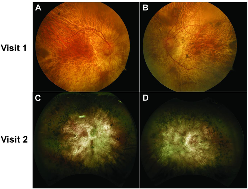
Figure 1.
Color fundus (top row) and widefield retina photography (bottom row) images showing widespread areas of chorioretinal atrophy and exposed underlying large choroidal vessels on both the right (A, C) and left (B, D) eye at visits 1 and 2. An island of spared retina on the parafoveal region was also observed bilaterally at both visits. Extensive intraretinal pigment migration in the periphery can be seen clearly in the widefield fundus images (C, D). Despite the different imaging modalities used, no significant progression is observed between the first and second visits. The difference in color and exposure is attributed to the different imaging modalities, but an island of spared retina of similar shape and size can be appreciated on both eyes at both visits.

