Figure 2.

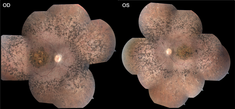
Color fundus photography of P2 reveals localized macular atrophy of the retinal pigment epithelium circumscribed by a confluent bone spicule pigment pattern in the beyond the vascular arcades in the periphery in both eyes.

Color fundus photography of P2 reveals localized macular atrophy of the retinal pigment epithelium circumscribed by a confluent bone spicule pigment pattern in the beyond the vascular arcades in the periphery in both eyes.