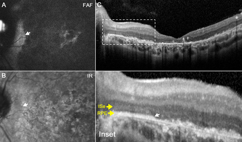
Figure 3.

Scanning laser ophthalmoscopy images from the 21-year-old sister (P2). Fundus autofluorescence and infrared images reveal macula-wide hypoautofluorescence (a) and hyporeflectance (b), respectively, with peripapillary sparing in each eye. Spectral domain-optical coherence tomography reveals loss of photoreceptors and ellipsoid nuclei in a similar pattern (C). Magnification reveals intact retinal architecture limited to the peripapillary region (inset).
