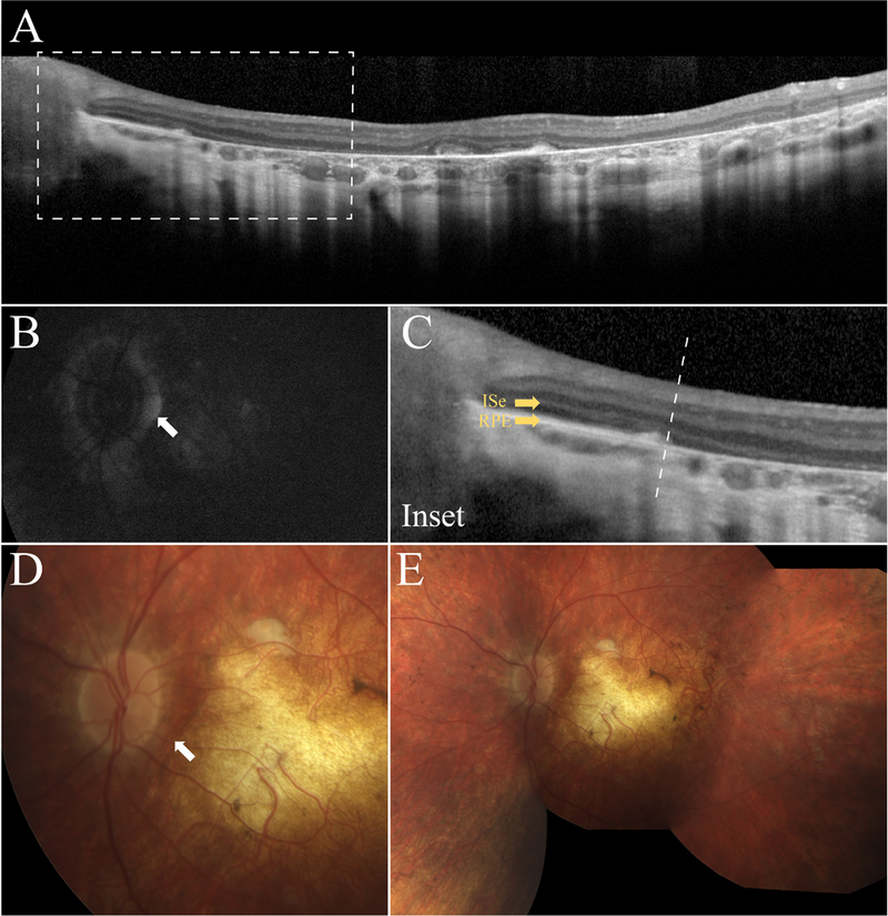
Figure 4.

Retinal imaging of the 10-year old boy (P4). SD-OCT shows extensive RPE and photoreceptor atrophy in the macula (A) and relative preservation of the RPE and retinal structure in the peripapillary region (C). SW-AF (B) imaging and color fundus photography (D and E, respectively) corroborate SD-OCT findings and show ubiquitous hypoautoflourescence and hyporeflectance that is spared immediately surrounding the optic nerve.
