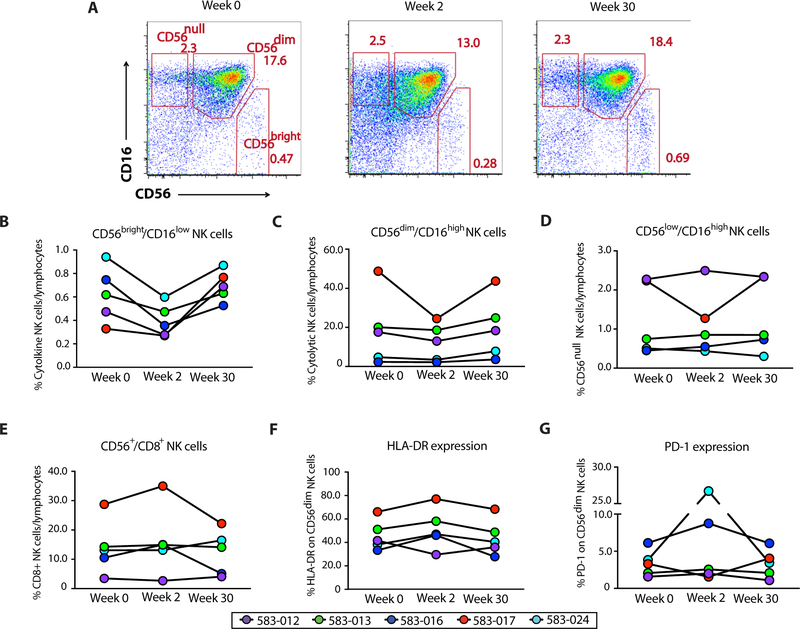
Fig. 6. Anti-α4β7 therapy results in the activation of circulating NK cell subsets.
(A) NK cell phenotype at weeks 0, 2, and 30 of therapy with VDZ. After exclusion of dead cells, monocytes, B cells, and T cells, cytolytic NK cells were gated as CD56dimCD16high NK cells, cytokine-secreting NK cells were gated as CD56brightCD16low NK cells, and CD56null cells were gated as CD56lowCD16high NK cells. (B to E) Composite graphs representing frequency of cytokine-producing (B), cytolytic (C), and CD56null NK cells (D), as well as CD56+CD8+ NK cells (E) from each of the five subjects (color-coded) at weeks 0, 2, and 30 of therapy with VDZ. (F and G) Change in the expression of HLA-DR (F) and PD-1 (G) on cytolytic CD56dimCD16high NK cells from five subjects (color-coded) at weeks 0, 2, and 30 of therapy with VDZ.

