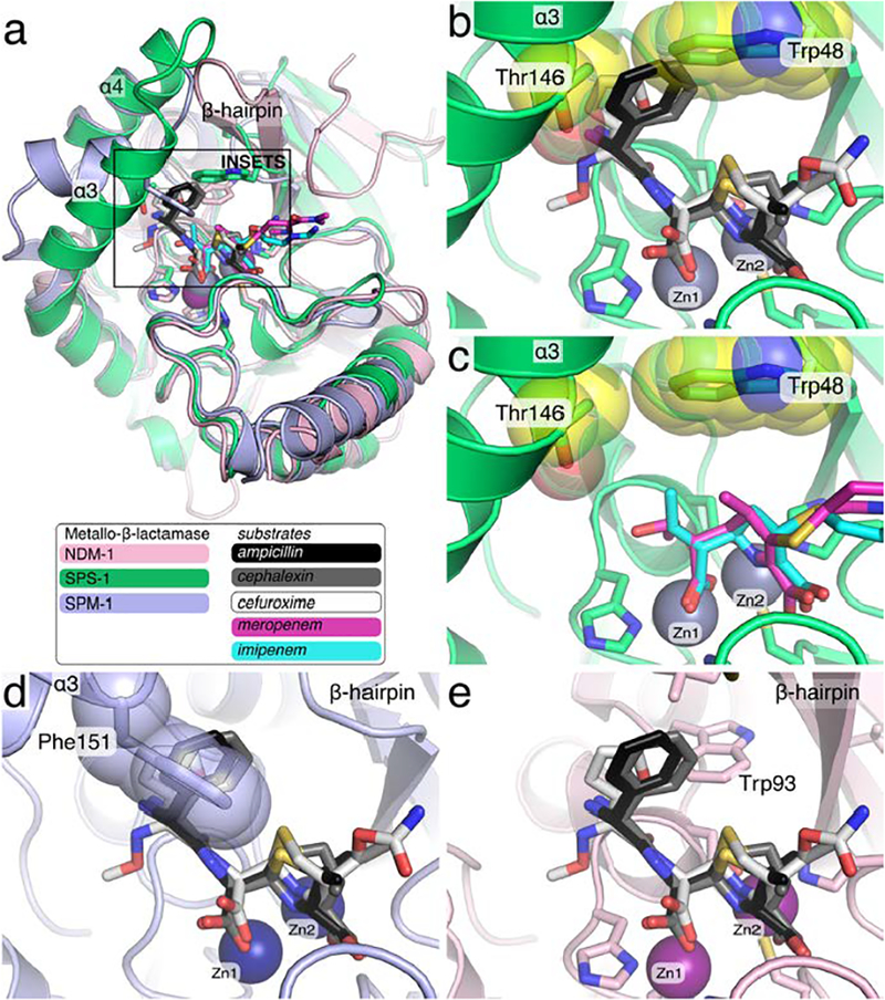
Figure 9.

Overlay of S. smaragdinae SPS-1 and SPM-1 onto NDM-1 complexes with hydrolyzed antibiotics. The structures of SPS-1 (green, PDB ID 6CQS) and SPM-1 (light blue, PDB ID 4BP0) were superimposed onto NDM-1 (pink, PDB ID 3Q6X) with SPS-1 Zn(II) ions (grey), SPM-1 Zn(II) ions (navy blue), and NDM-1 Zn(II) ions (purple) shown as spheres. The square in (a) shows the zoom used for insets b-e.Hydrolyzed antibiotics solved in complex with NDM-1 are shown as sticks including imipenem (cyan, PDB ID 5YPL), meropenem (magenta, 5YPK), ampicillin (black, PDB ID 3Q6X), cephalexin (grey, PDB ID 4RL2), and cefuroxime (white, PDB ID 4RL0). The side chains of SPS-1 residues Thr146 and Trp48 (yellow spheres) clash with ampicillin, cephalexin, and cefuroxime.
