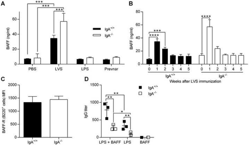
Fig. 6.
The role of BAFF in IgG responses to polysaccharide antigens. IgA+/+ and IgA−/− mice were immunized with PBS, F. tularensis LVS, F. tularensis LPS or Prevnar pneumococcal vaccine. (A) On day 7, BAFF levels in serum were determined by ELISA. (B) Kinetics of BAFF serum levels over a 5 weeks period following live LVS immunization. (C) Median fluorescence intensity (MFI) of BAFF receptor expression on B220+ cells from naïve mice. Splenocytes (106 cell/well) from naïve mice were co-cultured with LVS LPS (5 μg/ml), BAFF (25 ng/ml) or LPS + BAFF for 5 days. (D) Culture supernatants were analyzed for total IgG levels by ELISA. The data represent the means of 3 to 4 mice per group ± SD. *, P < 0.05; **, P < 0.01; ***, P<0.001 as analyzed by an ANOVA analysis, followed by Bonferroni’s multiple comparisons test.

