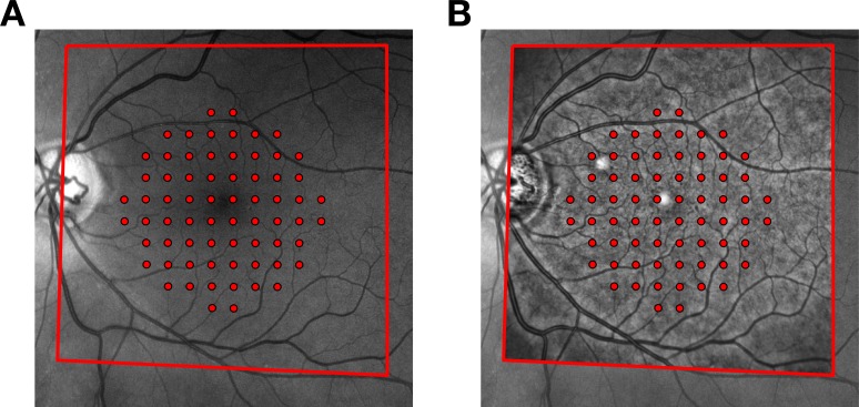
Figure 1.
Matching of fundus images from the Compass perimeter and the Spectralis SD-OCT. (A) Exemplar fundus image of a glaucoma patient with the locations of the 10-2 grid superimposed, as recorded by the tracking system. The red outline indicates the fundus area that was matched with the image from the Spectralis. (B) Matched image from the Spectralis distorted using a projective transformation and superimposed to the fundus image from Compass. The transformation establishes a two-way relationship between the structural map produced by the OCT and the functional map produced by the perimeter. As such, it can be used to map the tested locations from Compass onto the macular OCT map and vice versa.

