Fig. 2.
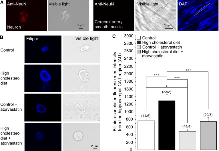
High-cholesterol diet and atorvastatin therapy modulate neuronal cholesterol content in the CA1 hippocampal region. A: Validation of the neuronal origin of the cellular content in the CA1 hippocampal region using neuron-specific immunostaining followed by confocal microscopy imaging. Immunofluorescence staining of neuronal tissue with an anti-NeuN Ab resulted in a fluorescence signal (leftmost snapshot). This staining failed to yield a signal from rat cerebral artery vasculature (three snapshots on the right). For cerebral artery vasculature, the silhouette of an individual myocyte is highlighted in the visible light spectrum and in the DAPI-stained specimen (rightmost snapshot). The sharp fluorescence image of the myocyte nucleus confirms that the vasculature was in the focal plane during imaging. B: Original representative snapshots showing filipin staining of isolated neuronal cells from the CA1 hippocampal brain region of rats on control diet, high-cholesterol diet, control diet supplemented by atorvastatin, and high-cholesterol diet supplemented by atorvastatin. C: Averaged data of filipin-associated fluorescence intensity from the hippocampal CA1 region of rats on control diet, high-cholesterol diet, and high-cholesterol diet supplemented by atorvastatin. AU, arbitrary units. (n/N) is the number of cells/number of animal donors. Statistically significant difference is indicated. * P < 0.05; *** P < 0.001.

