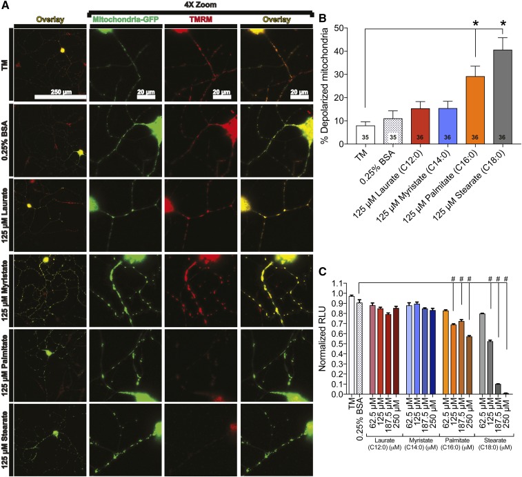
Fig. 5.
LCSFAs palmitate and stearate induce mitochondrial depolarization in mouse DRG neurons. A: DRG neurons expressing mito-GFP (green) were evaluated for changes in TMRM staining (red) using an overlay (yellow) of mito-GFP and TMRM signal that appears yellow in polarized mitochondria (left overlay column). Each neuron is displayed with a 4× zoom to depict the mitochondria-GFP signal, TMRM signal, and the merged image (right overlay column). Treatment conditions that retained DRG neuron mitochondrial membrane potential include TM, 0.25% BSA, 125 μM laurate, and 125 μM myristate. The 125 μM palmitate and stearate treatments exhibit diffuse TMRM staining (bottom two rows). B: Quantitation of TMRM signal in DRG neurons treated with 125 μM laurate (red bar) and myristate (blue bar) showed no significant effect on mitochondrial membrane potential relative to the TM and 0.25% BSA controls. Palmitate (orange bar) and stearate (black bar) at a concentration of 125 μM, however, exhibited a significant increase in the percentage of depolarized mitochondria relative to 125 μM laurate, 125 μM myristate, and the TM and 0.25% BSA control-treated neurons. The number displayed in the bar is the total number of DRG neurons that were evaluated for each treatment condition in three separate experimental trials. C: Palmitate- and stearate-treated 50B11 DRG neurons (from 125 to 250 μM) exhibit a reduction in intracellular ATP level compared with the 0.25% BSA control. Laurate and myristate treatments had no effect on ATP level. Values are expressed as mean ± SEM. * P < 0.01, ordinary one-way ANOVA with Tukey’s multiple-comparisons test (B); #P < 0.0001, ordinary one-way ANOVA with Tukey’s multiple-comparisons test (C).

