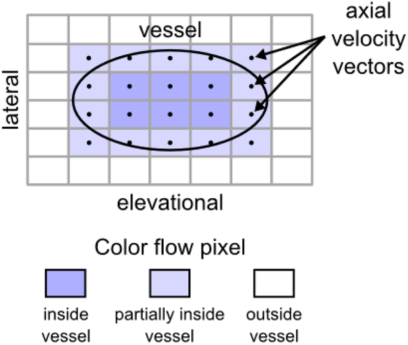
Figure 2:
Power-weighted surface integration of Doppler-measured velocity vectors (SIVV) illustrated on a vessel with an ellipsoidal cross section (black outline) in the lateral-elevational imaging surface (c-surface). Each color flow pixel that intersects the vessel possesses a Doppler-measured axial velocity vector that is normal to the area element. Axial velocity vectors point out of the page. Color flow pixels positioned inside the vessel correspond to 100% blood, those outside the vessel correspond to 0% blood, and those partially inside the vessel correspond to a value between 0% and 100% blood. Integration of flow over the c-surface yields volume flow through the shunt.

