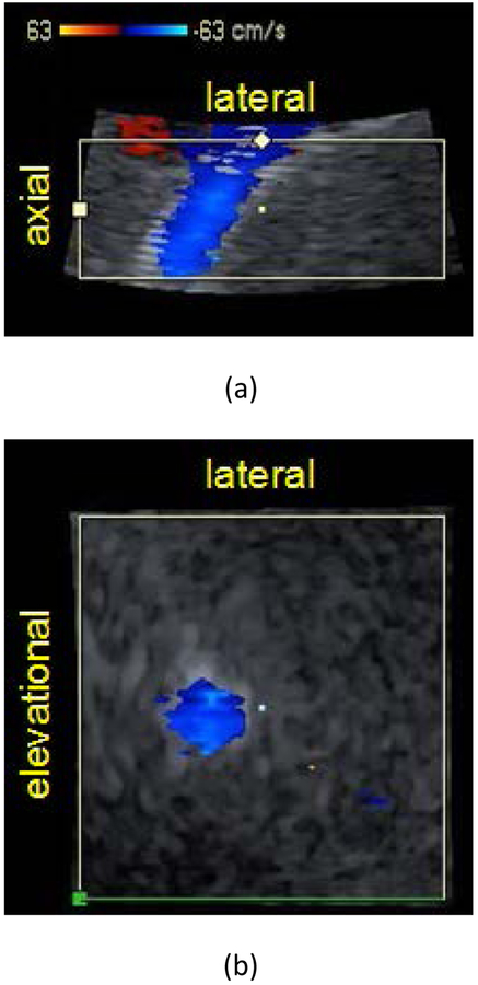
Figure 3:
Color flow images of transjugular intrahepatic portosystemic shunt (TIPS) in (a) axial-lateral and (b) elevational-lateral (c-surface) views for patient 4 post revision. Panels (a) and (b) coincide at the center point marked in each view. Color flow focus depth is 11.25 cm and is positioned at the axial center of (a). The elevational-lateral surface intersects the shunt at a depth of 11.25 cm. B-Mode image striations in (a) represent stent mesh boundary. Color bar indicates velocity in cm/s.

