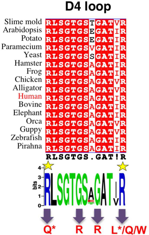
Figure 5. Sequences of the D4 loop of phosphoglucomutase in diverse eukaryotic organisms.
Top: a multiple sequence alignment (spanning residues 503 to 515 of human PGM) highlighting identical residues with red background. Bottom: A consensus Web Logo (Crooks et al., 2004) of the D4 loop. R503 and R515 are highlighted by yellow star; variants relevant to this study are indicated with arrows at bottom. Those with confirmed roles in PGM1 deficiency are marked with an asterisk.

