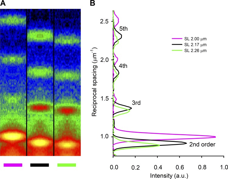
Figure 2.
X-ray sarcomere reflections recorded with the detector at 31 m from the preparation. (A) Meridional slices of 2-D diffraction patterns from a trabecula in diastole at three different lengths as indicated by the color code (magenta, 2.81 mm; black, 3.19 mm; green, 3.40 mm; corresponding SLs indicated with the same color code in the inset in B), showing the orders of the sarcomere reflections from second to fifth. At this camera length, the first order is masked by the beam stop. Total exposure time, 1.8 ms for each pattern. (B) Meridional intensity profiles from axial integration of the 2-D patterns in A. The lines and the SLs reported in the inset have the same color code as the corresponding trabecula lengths in A. a.u., arbitrary units.

