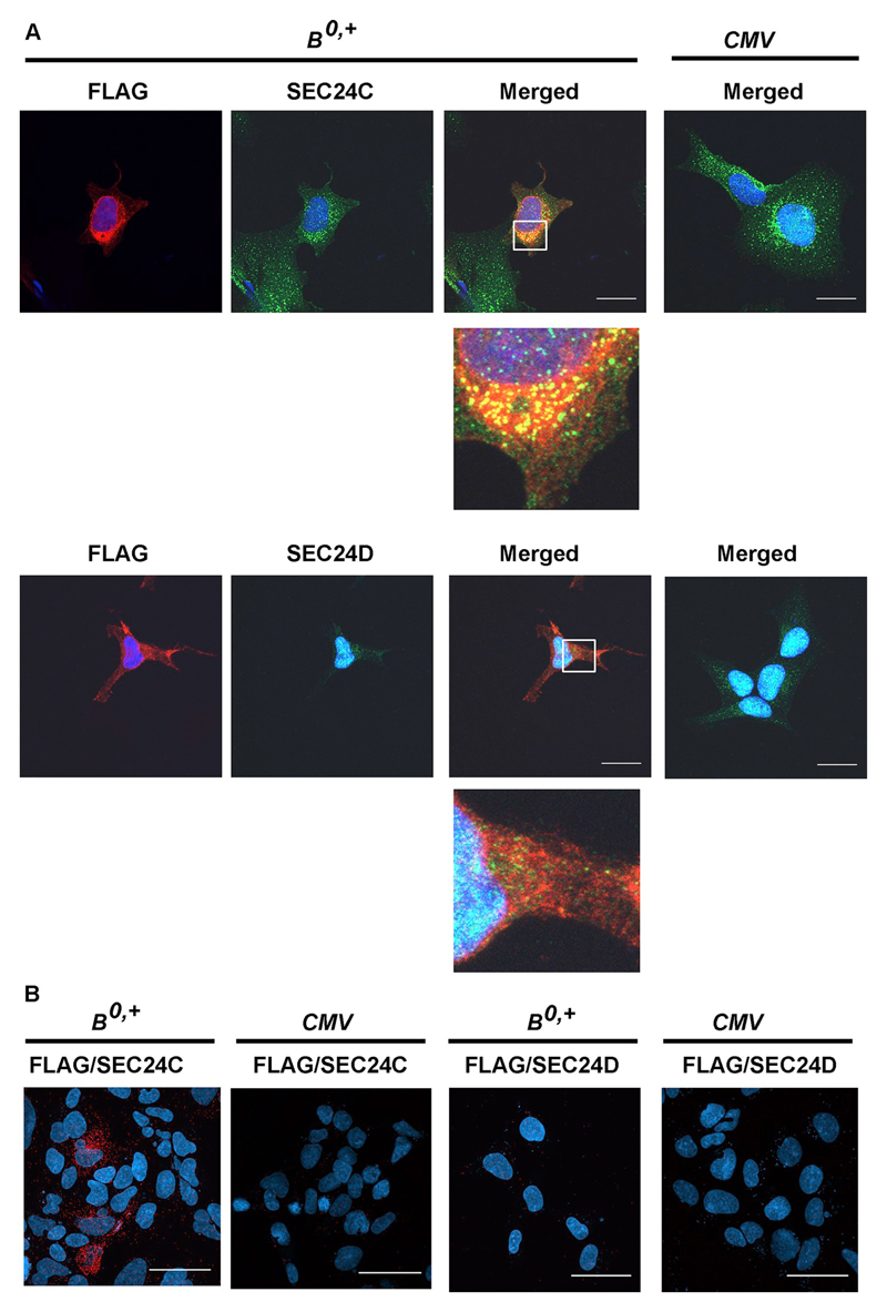
Fig. 4.
Analysis of ATB0,+ interaction with SEC24C and SEC24D. HEK293 cells were transfected for 48 h with the p3xFLAG-CMV14/B0,+ vector (B0,+) or p3xFLAG-CMV14 (CMV). (A) Localization of ATB0,+ was detected with anti-FLAG antibody (red), SEC24C and SEC24D with the corresponding antibodies (green), nuclei with DAPI (blue). Magnifications of selected areas are shown below the merged pictures. Bar 20 μm. (B) Proximity ligation assay performed with anti-FLAG and either anti-SEC24C or anti-SEC24D antibodies. Representative pictures out of 10 different images are shown. Bar 50 μm.

