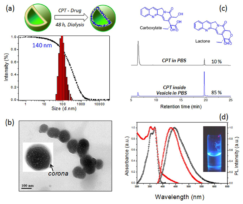
Figure 5.
Schematic representation of the encapsulation of camptothecin in the vesicle and its DLS histograms (a). HR-TEM image of the CPT loaded vesicles (b). The inset showed the expanded image of the vesicles with well-defined corona with respect to vesicular assemblies. HPLC traces of CPT in PBS and encapsulated in the vesicle in the carboxylic and lactone forms (c). Absorbance and fluorescence spectra of CPT loaded vesicles in PBS (d).

