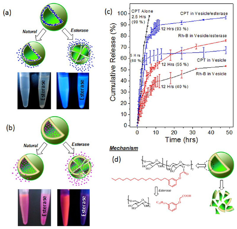
Figure 6.
Schematic drug release of vesicular scaffolds under normal dialysis and in the presence of esterase enzymes. The photographs of the vials showed the formation of precipitate over a prolonged storage of the CPT loaded (a) and Rh-B loaded vesicles (b) in the presence of esterase. The left and right of the vials are corresponding to the Rh-B or CPT loaded vesicles in the absence or presence of esterase, respectively. Cumulative drug release of CPT, CPT loaded vesicles, and Rh-B loaded vesicles (c). The schematic model represents the disassociation of vesicular scaffolds in the presence of esterase (d).

