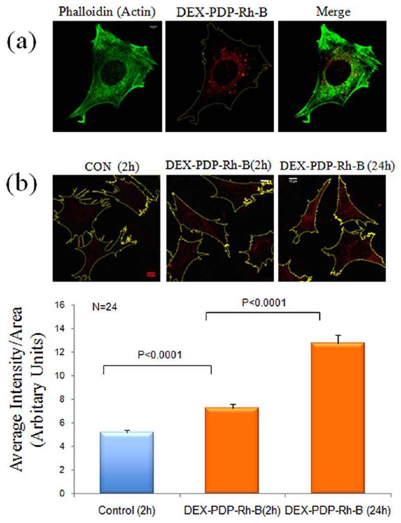
Figure 8.
Endocytosis of DEX-PDP-Rh-B in mouse fibroblasts. (a) Endocytosed DEX-PDP-Rh-B shows a distinct perinuclear localization in mouse fibroblasts. Actin cytoskeletal network in cells is stained with phalloidin. (b) Fluorescence confocal images of control untreated cells and cells incubated with DEX-PDP-Rh-B for 2 and 24 h were recordered, their spread area mapped, and fluorescence intensity in the cell area analyzed using the Image J densitometric software. Mean of fluorescence intensities is represented in the graph.

