Figure 1.
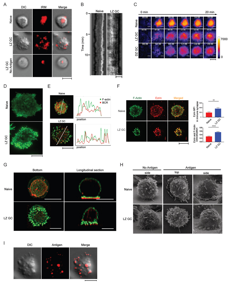
GC B Cells Engage Antigen through BCRs Concentrated in F-Actin and Ezrin-Rich Pod-like Structures. (A) DIC, IRM, and merged images of immune synapse of live naïve B cells placed on antigen-containing PLB and live LZ GC B cells placed on antigen-containing PLB or PLB with no antigen. (B) Kymographs of DIC images of naïve and LZ GC B cell immune synapse on antigen-containing PLB. (C) Membrane movement in the immune synapses of live naïve, LZ GC, and DZ GC B cells with time on antigen-containing PLB imaged by IRM. (D) STED super-resolution images of F-actin formed in immune synapses of naïve B cells and GC B cells placed on PLB that contained antigen. (E) Colocalization of F-actin and BCR in immune synapses of naïve and LZ GC B cells imaged by confocal microscopy on PLB that contained antigen. (F) (Left panel) Confocal microscopy images of immune synapses of naïve B cells and LZ GC B cells on antigen-coated PLB stained with Alexa Fluor 488 phalloidin for F-actin (green) and antibodies specific for ezrin (red). (Top right panel) Quantification of the MFI of ezrin and (Bottom right panel) colocalization of ezrin with F-actin (bottom) in the immune synapse of confocal images. (G) Bottom and orthogonal views of F-actin (green) and BCR (red) in naïve and LZ GC B cells imaged by confocal microscopy on antigen-containing PLB. (H) Side and top views of naïve and LZ GC B cells imaged by SEM on PLB without or with antigen. (I) Colocalization of pod-like structures and antigens in immune synapse of LZ GC B cells imaged by TIRFM on antigen-containing PLB. Scale bars are 5µm. ns>0.05, *P≤0.05, **P≤0.01, ***P≤0.001, and ****P≤0.0001 (unpaired t-test). Data are representative of two experiments (F: mean and s.e.m.).

