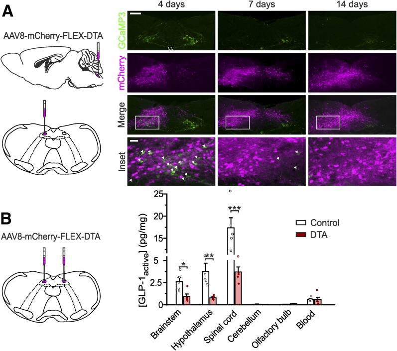
Figure 2.
NTS PPG neurons are the main source of brain GLP-1. A: Expression of mCherry and GCaMP3 (as a marker for PPG neurons) 4, 7, and 14 days after unilateral stereotaxic injection of AAV8-mCherry-FLEX-DTA into the NTS of a Glu-Cre/GCaMP3 mouse (schematic on left). White arrows indicate the remaining GCaMP3-positive PPG neurons. Scale bars: top panels, 100 μm; inset, 20 μm. B: Protein levels of active GLP-1 (normalized to total protein) detected in several brain regions after bilateral stereotaxic injection of AAV8-mCherry-FLEX-DTA or a control virus (AAV1/2-FLEX-Perceval). Brainstem P = 0.0317; hypothalamus P = 0.0079 (Mann-Whitney U test); spinal cord P = 0.0004 (unpaired t test). Data are given as the mean ± SEM; n = 5 in each group. *P < 0.05, **P < 0.01, ***P < 0.001.

